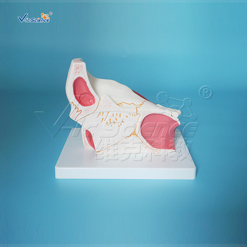
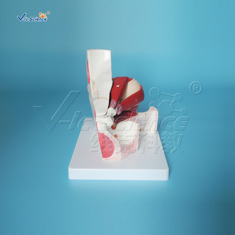
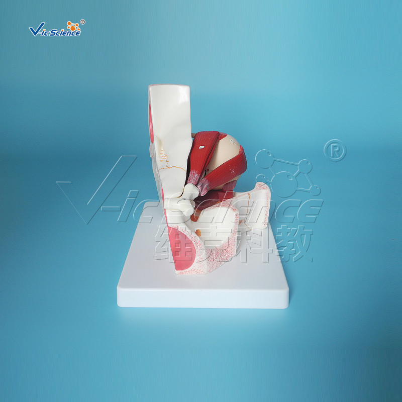
|
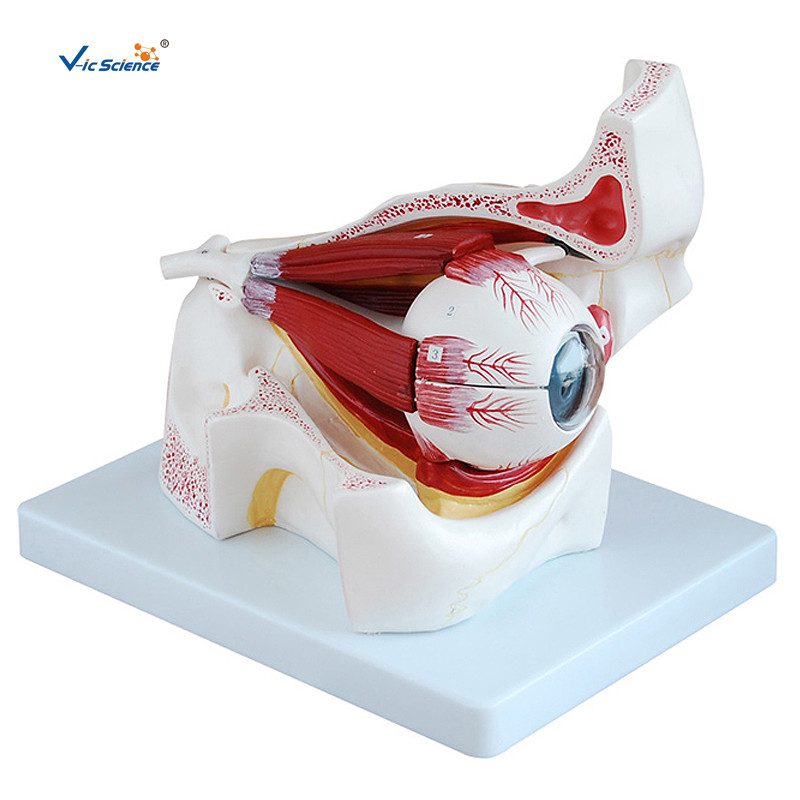
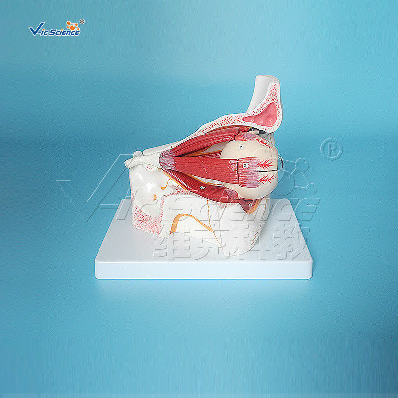
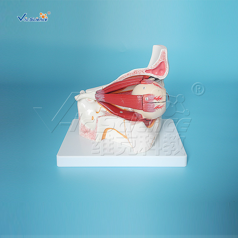
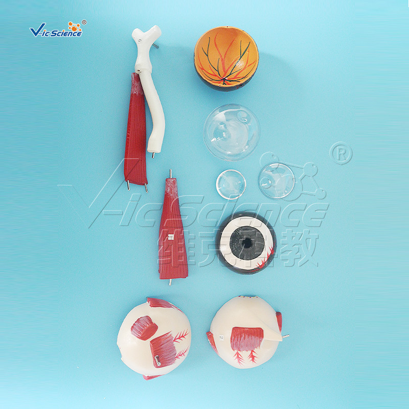
There are two eyes, situated on the left and the right of the face. Ada dua mata, terletak di kiri dan kanan wajah. They sit in two bony cavities called the orbits, which are present in the skull. Mereka duduk di dua rongga tulang yang disebut orbit, yang ada di tengkorak. Six extraocular muscles attach directly to the eyes to assist with movement. Enam otot ekstraokular menempel langsung ke mata untuk membantu gerakan. The front visible part of the eye is made up of the whitish sclera, a coloured iris, and the pupil. Bagian depan mata yang terlihat terdiri dari sklera keputihan, iris berwarna, dan pupil. A thin layer called the conjunctiva sits on top of this. Lapisan tipis yang disebut konjungtiva duduk di atas ini. The front part is also called the anterior segment of the eye. Bagian depan juga disebut segmen anterior mata.
The eye is not shaped like a perfect sphere, rather it is a fused two-piece unit, composed of a anterior (front) segment and the posterior (back) segment. Mata tidak berbentuk seperti bola sempurna, melainkan merupakan unit dua potong yang menyatu, terdiri dari segmen anterior (depan) dan segmen posterior (belakang). The anterior segment is made up of the cornea, iris and lens. Segmen anterior terdiri dari kornea, iris dan lensa. The cornea is transparent and more curved, and is linked to the larger posterior segment, composed of the vitreous, retina, choroid and the outer white shell called the sclera. Kornea transparan dan lebih melengkung, dan terkait dengan segmen posterior yang lebih besar, terdiri dari vitreous, retina, koroid dan kulit putih luar yang disebut sklera. The cornea is typically about 11.5 mm (0.3 in) in diameter, and 0.5 mm (500 μm) in thickness near its center. Kornea biasanya berdiameter sekitar 11,5 mm (0,3 in.), Dan ketebalan 0,5 mm (500 m) di dekat pusatnya. The posterior chamber constitutes the remaining five-sixths; Ruang posterior membentuk sisa lima perenam; its diameter is typically about 24 mm. diameternya biasanya sekitar 24 mm. The cornea and sclera are connected by an area termed the limbus. Kornea dan sklera dihubungkan oleh area yang disebut limbus. The iris is the pigmented circular structure concentrically surrounding the center of the eye, the pupil, which appears to be black. Iris adalah struktur lingkaran berpigmen yang secara konsentris mengelilingi pusat mata, pupil, yang tampak berwarna hitam. The size of the pupil, which controls the amount of light entering the eye, is adjusted by the iris' dilator and sphincter muscles. Ukuran pupil, yang mengontrol jumlah cahaya yang masuk ke mata, disesuaikan dengan dilator iris dan otot sfingter.
Light energy enters the eye through the cornea, through the pupil and then through the lens. Energi cahaya memasuki mata melalui kornea, melalui pupil dan kemudian melalui lensa. The lens shape is changed for near focus (accommodation) and is controlled by the ciliary muscle. Bentuk lensa diubah untuk fokus dekat (akomodasi) dan dikendalikan oleh otot ciliary. Photons of light falling on the light-sensitive cells of the retina (photoreceptor cones and rods) are converted into electrical signals that are transmitted to the brain by the optic nerve and interpreted as sight and vision. Foton cahaya yang jatuh pada sel peka cahaya retina (kerucut fotoreseptor dan batang) diubah menjadi sinyal listrik yang ditransmisikan ke otak oleh saraf optik dan diartikan sebagai penglihatan dan penglihatan.
|
 Pesan Anda harus antara 20-3.000 karakter!